


The Most Downloaded BMJ Article Still Fascinates 20 Years Later
Hint: It’s about sex

If you enjoyed this article, please share it with a friend or consider becoming a paid subscriber. Weekday articles are free. Sunday’s article is for paid subscribers only.
The idea started, like all great ideas do, with a question: What is happening inside a couple’s body while they are having sex? In the early 1990s, a Dutch team led by gynecologist Willibrord Weijmar Schultz and physiologist Pek van Andel decided to find out.
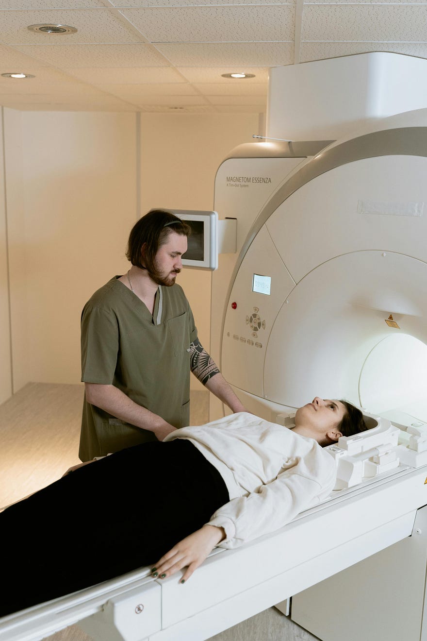
So they stuffed some brave volunteers into a magnetic resonance imaging (MRI) and took a picture mid coitus. Could MRI — that big, noisy donut usually reserved for brain tumors and torn ACLs — clear up myths about what our plumbing is up to during sex?
The question was simple. The methodology…well, let’s just say it got some pushback. Funding was scarce. Permission was even scarcer. Imagine trying to explain to the ethics board why a 1.5 Tesla magnet would be cradling naked, entwined bodies on a Saturday. Awkward.
Still, the curiosity was warranted. Until then, most anatomical knowledge about intercourse was cobbled together from guesswork, glass tubes, and, frankly, a lot of imagination.
In 1493, Leonardo drew what he thought intercourse looked like: semen falling through the man’s spine like a Rube Goldberg machine and the woman’s breast ducts connected directly to her womb like some dairy farm plumbing system. Unfortunately, Leonardo got a lot wrong, especially with the shape of the penis during sex.
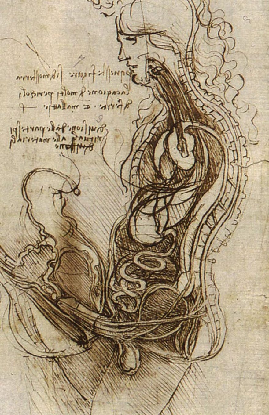
Leonardo wasn’t an idiot. He was just, like most of us, working with a lot of guesswork and no good lighting.
Later anatomists improved the lighting but not the theories. In the 1930s, gynecologist R. L. Dickinson used a glass tube, a vibrator, and a prayer to model what he thought the human body did during arousal. Masters and Johnson in the 1960s got closer but still relied on bimanual palpation — scientific code for poking around with fingers and squinting meaningfully.
Modern researchers also hit a few snafus. Sex, it turns out, is a lot less sexy when performed inside a glorified concrete torpedo tube with your pelvis pressed against cold metal, all while a disembodied voice cackles through the intercom: “Hold very still, please!”
Yet despite the long odds — and a few cases of unfortunate performance anxiety — the researchers persisted. Between 1991 and 1999, they ran thirteen experiments with eight couples and three intrepid solo women. At first, the images were blurry, courtesy of the ungainly early MRI machines. Then, in 1998, a pharmaceutical miracle named sildenafil (Viagra to you and me) swooped in. One man popped a tablet, climbed into the scanner, and delivered a performance long enough to yield crisp, scandalously clear images of coitus in progress.
When the team submitted their work to the prestigious British Medical Journal (BMJ) in 1999, no one quite knew what to make of it. It wasn’t exactly curing cancer, but it was the first time anyone had gotten such an intimate view of sex. “A striking image using a new technology,” wrote editor Tony Delamothe with the kind of bemused understatement one uses to describe a nephew’s garage band. Everyone agreed that readers might be interested in seeing it.
Interested, indeed.
As Delamothe later recounted, the paper became the most visited article in the history of the BMJ, outpacing two centuries’ worth of papers on measles outbreaks and heart transplants. It even won an Ig Nobel Prize in 2000 — an award for scientific achievements that “first make people laugh, then think.”
But what the researchers captured wasn’t just an anatomical curiosity. What they captured is a map to understanding why some sex positions feel so good and others…meh.
Keep reading with a 7-day free trial
Subscribe to Conversations with Carlyn to keep reading this post and get 7 days of free access to the full post archives.